
Coronary Angiogram

A coronary angiogram is a procedure that uses X-ray imaging to see your heart's blood vessels. The test is generally done to see if there's a restriction in blood flow going to the heart.
Coronary angiograms are part of a general group of procedures known as heart (cardiac) catheterizations. Cardiac catheterization procedures can both diagnose and treat heart and blood vessel conditions. A coronary angiogram, which can help diagnose heart conditions, is the most common type of cardiac catheterization procedure.
During a coronary angiogram, a type of dye that's visible by an X-ray machine is injected into the blood vessels of your heart. The X-ray machine rapidly takes a series of images (angiograms), offering a look at your blood vessels. If necessary, your doctor can open clogged heart arteries (angioplasty) during your coronary angiogram.

Why it's done

Your doctor may recommend that you have a coronary angiogram if you have:
·         Symptoms of coronary artery disease, such as chest pain (angina)
·         Pain in your chest, jaw, neck or arm that can't be explained by other tests
·         New or increasing chest pain (unstable angina)
·         A heart defect you were born with (congenital heart disease)
·         Abnormal results on a noninvasive heart stress test
·         Other blood vessel problems or a chest injury
·         A heart valve problem that requires surgery
Because there's a small risk of complications, angiograms aren't usually done until after noninvasive heart tests have been performed, such as an electrocardiogram, an echocardiogram or a stress test.

Risks

As with most procedures done on your heart and blood vessels, a coronary angiogram has some risks, such as radiation exposure from the X-rays used. Major complications are rare, though. Potential risks and complications include:
·         Heart attack
·         Stroke
·         Injury to the catheterized artery
·         Irregular heart rhythms (arrhythmias)
·         Allergic reactions to the dye or medications used during the procedure
·         Kidney damage
·         Excessive bleeding
·         Infection

How you prepare

In some cases, coronary angiograms are performed on an emergency basis. More commonly, though, they're scheduled in advance, giving you time to prepare.
Angiograms are performed in the catheterization (cath) lab of a hospital. Your health care team will give you specific instructions and talk to you about any medications you take. General guidelines include:
·         Don't eat or drink anything after midnight before your angiogram.
·         Take all your medications to the hospital with you in their original bottles. Ask your doctor about whether or not to take your usual morning medications.
·         If you have diabetes, ask your doctor if you should take insulin or other oral medications before your angiogram.

What you can expect

Before the procedure

Before your angiogram procedure starts, your health care team will review your medical history, including allergies and medications you take. The team may perform a physical exam and check your vital signs — blood pressure and pulse.
You'll also empty your bladder and change into a hospital gown. You may have to remove contact lenses, eyeglasses, jewelry and hairpins.

During the procedure

·     Cardiac catheterization procedure approaches
For the procedure, you lie on your back on an X-ray table. Because the table may be tilted during the procedure, safety straps may be fastened across your chest and legs. X-ray cameras may move over and around your head and chest to take pictures from many angles.
An IV line is inserted into a vein in your arm. You may be given a sedative through the IV to help you relax, as well as other medications and fluids. You'll be very sleepy and may drift off to sleep during the procedure, but you'll still be able to be easily awakened to follow any instructions.
Electrodes on your chest monitor your heart throughout the procedure. A blood pressure cuff tracks your blood pressure and another device, a pulse oximeter, measures the amount of oxygen in your blood.
A small amount of hair may be shaved from your groin or arm where a flexible tube (catheter) will be inserted. The area is washed and disinfected and then numbed with an injection of local anesthetic.
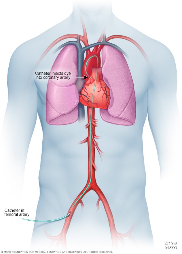
·        

Coronary angiogram

A small incision is made at the entry site, and a short plastic tube (sheath) is inserted into your artery. The catheter is inserted through the sheath into your blood vessel and carefully threaded to your heart or coronary arteries.
Threading the catheter shouldn't cause pain, and you shouldn't feel it moving through your body. Tell your health care team if you have any discomfort.
Dye (contrast material) is injected through the catheter. When this happens, you may have a brief sensation of flushing or warmth. But again, tell your health care team if you feel pain or discomfort.
The dye is easy to see on X-ray images. As it moves through your blood vessels, your doctor can observe its flow and identify any blockages or constricted areas. Depending on what your doctor discovers during your angiogram, you may have additional catheter procedures at the same time, such as a balloon angioplasty or a stent placement to open up a narrowed artery.
Having an angiogram takes about one hour, although it may be longer, especially if combined with other cardiac catheterization procedures. Preparation and post-procedure care can add more time.

After the procedure

When the angiogram is over, the catheter is removed from your arm or groin and the incision is closed with manual pressure, a clamp or a small plug.
You'll be taken to a recovery area for observation and monitoring. When your condition is stable, you return to your own room, where you're monitored regularly.
You'll need to lie flat for several hours to avoid bleeding if the catheter was inserted in the groin. During this time, pressure may be applied to the incision to prevent bleeding and promote healing.
You may be able to go home the same day, or you may have to remain in the hospital overnight. Drink plenty of fluids to help flush the dye from your body. If you're feeling up to it, have something to eat.

Results

An angiogram can show doctors what's wrong with your blood vessels. It can:
·         Show how many of your coronary arteries are blocked or narrowed by fatty plaques (atherosclerosis)
·         Pinpoint where blockages are located in your blood vessels
·         Show how much blood flow is blocked through your blood vessels
·         Check the results of previous coronary bypass surgery
·         Check the blood flow through your heart and blood vessels

Knowing this information can help your doctor determine what treatment is best for you and how much danger your heart condition poses to your health. Based on your results, your doctor may decide, for instance, that you would benefit from having coronary angioplasty or stenting to help clear clogged arteries. It's also possible that angioplasty or stenting could be done during your angiogram to avoid needing another procedure

Comments

Popular posts from this blog

Vascular Catheterization - Coding

Vascular system anatomy and terminology to make reporting these procedures less challenging. A thorough understanding of the anatomy and medical terminology of the vascular system is required to accurately code arterial vessel procedures. Here’s a quick run-through of clinical information and coding guidance to show you how order affects coding. Order Matters Please refer to Appendix L of the CPT® code book for the vascular families. The aorta is the major artery. Each main vessel branching off the aorta is a first order vessel (e.g., left common carotid). First order vessels have several vessels branching off them, designated as second order vessels (e.g., right subclavian and axillary). Second order vessels have several vessels branching off them, designated as third order vessels (e.g., right internal carotid). Third order vessels have several smaller vessels branching off them, designated as beyond the third order (e.g., left deep palmar arch). Catheterization Can Ei...

Injections & Infusions Coding

Here are the three basic coding rules that you must adhere to when billing for Injection/Infusion services: Rule #1: Pay Attention to the Administration Route When you’re looking at the treatment note, you can first look for hints to proper coding by identifying the Route of Administration.  the Injection/Infusion CPT® codes fall into one of three major categories: 1. Intravenous Infusions (IV) 2. Intravenous Pushes (IP) 3. Injections (Sub-Q, IM) Don’t forget: The Centers for Medicare & Medicaid Services (CMS) regulations look at the way a drug is administered only as “the physical process by which the drug enters the patient’s body” – not whether a medical professional administers the drug or supervises the administration. But the administration route hierarchy isn’t the only one you need to understand for Injection/Infusion coding — there is another hierarchy for the type of agent that works in tandem with the administration one. Rule #2: Unde...

Systemic vs. Pulmonary Circulation

What Does the Heart Do? The heart is a pump, usually beating about 60 to 100 times per minute. With each heartbeat, the heart sends  blood  throughout our bodies, carrying oxygen to every cell. After delivering the oxygen, the blood returns to the heart. The heart then sends the blood to the  lungs  to pick up more oxygen. This cycle repeats over and over again. What Does the Circulatory System Do? The circulatory system is made up of blood vessels that carry blood away from and towards the heart.  Arteries  carry blood away from the heart and  veins  carry blood back to the heart. The circulatory system carries oxygen, nutrients, and  hormones  to cells, and removes waste products, like carbon dioxide. These roadways travel in one direction only, to keep things going where they should. What Are the Parts of the Heart? The heart has four chambers — two on top and two on bottom: ·       ...

Place Holder X

The letter " x ” serves as a placeholder when a code contains fewer than six characters and a seventh character applies. The " x ” also allows for future expansion of the codes. When reporting ICD - 10 - CM codes, coders must add a placeholder so the seventh character is in the correct position. Not every ICD-10-CM code with a seventh character has a sixth character—or even a fifth or fourth character for that matter. This frequently occurs with poisonings and injuries. The letter "x” serves as a placeholder when a code contains fewer than six characters and a seventh character applies. The "x” also allows for future expansion of the codes. When reporting ICD-10-CM codes, coders must add a placeholder so the seventh character is in the correct position. Without this placeholder to ensure characters appear in the correct positions, codes are invalid. For example, a patient presents with an accidental poisoning by an antiallergic drug. For the initial ...

Burns and Corrosions

Chemicals, such as lye or acid, can cause corrosion upon contact with a person's skin. As a professional coding specialist, you may need to code the diagnosis of a burn or corrosion. A chemical burn occurs when living tissue is exposed to a corrosive substance such as a strong acid or base. Chemical burns follow standard burn classification and may cause extensive tissue damage. Burns from a heat source are classified by depth (first, second, third, unspecified), extent, and agent. For multiple burns, sequence the highest degree burn first. Multiple burns of the same three-character category are coded to the highest degree. Non-healing burns are coded as acute burns. The extent of injury is best described using the percentage of the total body surface area (%TBSA) that is affected by a burn. The measurement of burn surface area is important during the initial management of people with burns for estimating fluid requirements and determining need for transfer to a burns service T...

Allograft vs Autograft vs Xenograft

Allo – Same species Auto – Same person Xeno – Animal (different species) An autologous transplant uses a person's own stem cells. ... In a reduced-intensity allogeneic transplant, doctors suppress the recipient's immune system enough so the donor stem cells can take root, or “engraft,” there. An autograft is a bone or tissue that is taken from a part of a person’s own body and transplanted into another. Often, surgeons will use a person’s own hamstring tendon to repair a damaged anterior cruciate ligament. Similarly, an autograft bone may be transplanted from a person’s hip to aide in a spinal fusion. Patients who undergo autograft procedures may experience increased postoperative pain from the second surgical (autograft) site. They may also require longer periods of rehabilitation. The use of allograft is advantageous because there is no second procedure required to remove and transfer a portion of the patient’s native bone or tissue. Surgical time may be...

Combination Codes

It’s one of the most important questions coders must ask while using ICD-10: Is there a single combination code that fully identifies the patient’s relevant conditions, or is it necessary to report two separate codes? The volume of combination codes in ICD-10 has increased, making it imperative for coders to be alert and aware of instances in which combination codes are applicable. Defining Combination Codes The ICD-10-CM Official Guidelines for Coding and Reporting describe combination codes as those used to classify the following: •         Two diagnoses •         A diagnosis with an associated secondary process (manifestation) •         A diagnosis with an associated complication Coders cannot — and should not — assign multiple diagnosis codes when a single combination code clearly identifies all aspects of the patient’s diagnosis. For example, say a pati...

Complete List of Modifiers

Modifier - as the name implies a modifier will modify a service / procedure or an item under certain circumstances for appropriate reimbursement. Modifiers may add information or change the description according to the physician documentation to give more specificity for the service or procedure rendered. Appending of an appropriate modifier will effectively respond to reimbursement. Modifier are two digit codes and are categorized into two levels 1. Level I Modifiers: Normally known as CPT Modifiers and consists of two numeric digits and are updated annually by AMA - American Medical Association. 2. Level II Modifiers: Normally known as HCPCS Modifiers and consists of two digits (Alpha / Alphanumeric characters) in the sequence AA through VP. These modifiers are annually updated by CMS - Centres for Medicare and Medicaid Services. Both the above levels of Modifiers are recognized nationally. List of Level I Modifiers: Modifier -21 Prolonged Evaluation and Man...

Spleen - Anatomy

The  spleen  is an organ located in the upper left abdomen, and is roughly the size of a clenched fist. In the adult, the spleen functions mainly as a blood filter, removing old red blood cells. It also plays a role in both cell-mediated and humoral immune responses. The spleen in located in the upper left quadrant of the abdomen, under cover of the diaphragm and  the ribcage – and therefore cannot normally be palpated on clinical examination (except when enlarged). It is an intraperitoneal organ, entirely surrounded by peritoneum (except at the splenic hilum). The spleen is connected to the stomach and kidney by parts of the greater omentum – a double fold of peritoneum that originates from the stomach: The spleen has a slightly  oval  shape. It is covered by a weak capsule that protects the organ whilst allowing it to expand in size. The outer surface of the spleen can be anatomically divided into two: ·      ...