Ischemic heart disease continues to be the leading cause of death in the United
States and worldwide (World Health Organization, 2017). It’s no surprise that
myocardial infarctions (MI) are frequently seen as reportable conditions when
coding inpatient encounters. Recent ICD-10-CM changes offer new codes to
further specify the type and cause of MIs.
How and where the myocardial death occurs determines the ICD-10-CM code
assignment.

MIs are categorized in several
ways. Historically, MIs were categorized based
on the thickness of the
myocardial necrosis. A transmural MI occurs when the
myocardial necrosis is full
thickness (extending from the endocardium through
the myocardium to the
epicardium), and a non-transmural MI includes necrosis
of the endocardium or the
endocardium and myocardium only.
Electrocardiogram findings are more commonly used
to identify the type of MI. This includes ST-elevation MIs (STEMI), non-ST-elevation
MIs (NSTEMI), Q-wave MIs, and non-Q-wave MIs.
An important change in the new
2018 ICD-10-CM Official
Guidelines for Coding
and Reporting is the addition of a code for an
unspecified AMI (I21.9). Previously, the unspecified AMI defaulted to
a STEMI (I21.3).
“Management practice guidelines
often distinguish between STEMI and non-
STEMI, as do many of the studies
on which recommendations are based,”
If the documentation of an
unspecified AMI defaults to a STEMI, but it is not treated as a STEMI, this could adversely
affect quality measures for clinical performance.
Specifically, based on the 2017
recommendations of the American Hospital
Association and the American
College of Cardiology Foundation, the established
goal of treatment for a patient
with a diagnosis of an acute STEMI is an elapsed time of 90 minutes or less from first
medical contact to primary percutaneous coronary intervention
(PCI) when presenting to a facility with PCI capabilities.
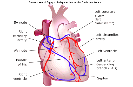
AMIs are further identified by
site, which corresponds with the coronary artery
involved (e.g., inferior wall
MI, anterior wall MI, etc.), physicians should document which
coronary artery was affected to capture the most specific code available. Documentation of a transmural MI
unspecified by site will code as I21.9, AMI unspecified, but when specified
by site or artery will code as a STEMI. Nontransmural MIs code as an NSTEMI.
The cause of an MI further
defines the type of MI and the ICD-10-CM code
assignment. As mentioned
earlier, the revised definition of a MI in 2007
emphasized the causes of the MI.
The classification of MIs was divided into five
types,
which is an important piece of the universal definition.
A type 1 MI is often referred to
as a “spontaneous” MI and is commonly associated with coronary artery disease
(CAD) and myocardial necrosis secondary to plaque rupture, erosion, dissection,
ulceration, or erosion that results in a thrombus in one or more of the coronary arteries
A type 2 MI occurs when
myocardial necrosis results from either a reduction in
oxygen supply or decreased blood
flow to the heart or an increase in the heart’s
need (demand) for oxygen.
According to Sandoval, Smith, Thordsen, & Apple
(2014), anemia, tachyarrythmia,
and respiratory failure were the most common
conditions predisposing patients
to a type 2 MI, and “it is anticipated that it [type 2 MIs] will be detected more
frequently once high sensitivity cardiac troponin assays are approved for clinical use in
the United States.”
Some other examples of increased
demand include severe aortic valve disease,
hypertension, and shock. One-fourth
of the patients observed with AMIs were diagnosed with a type 2 MI, and of
those, half had no significant CAD. Please note that it is a common error to
call out type 2 MIs as NSTEMI type 2. A type 2 MI is an AMI by definition and
not a NSTEMI
It was identified that “distinct
demographics, increased prevalence of multiple comorbidities, a high-risk
cardiovascular profile and an overall worse outcome” in patients diagnosed with
a type 2 MI compared to those diagnosed with a type 1 MI. Specifically, they
found patients diagnosed with type 2 MI to be older and female, and to have a
higher incidence of prior MIs, PCI or coronary artery bypass graft (CABG),
heart failure, chronic renal failure, and diabetes. “It is conceivable,” they
stated, “that elderly patients with multiple comorbidities and an underlying
coronary disease would be more susceptible to clinical changes that may
interfere with the delicate balance of myocardial supply and demand, ensuing in
type 2 MI”
Included in the universal
definition of MIs (but seen documented less frequently)
are type 3 MI, types 4a and 4b
MI, and type 5 MI. Type 3 MI refers to an AMI
when there is evidence of
myocardial ischemia based on ECG finding and/or a
new left bundle branch block,
but death occurs before cardiac biomarkers can be
obtained. Type 4a MI is an MI
occurring after PCI. Type 4b MI is associated with stent thrombosis
after PCI. Type 5 MI is associated with an MI after a CABG procedure
A final distinguishing factor
for coding purposes is the age of the MI. Subsequent MIs are MIs occurring within
four weeks or less of the initial MI. Code I22 is used for subsequent type 1 STEMI,
NSTEMI, and AMI, unspecified. A code for the initial MI
(I21.-) must be included.
The sequencing of these codes
depends on the reason for admission. If the
subsequent MI is identified as a
type 2 STEMI or NSTEMI, assign only I21.A1 and I21.A9 for type 3, 4, and 5 MIs.
MIs that occurred more than four weeks prior but are still being treated have the
appropriate aftercare code assigned, and those no longer
needing treatment are coded as I25.2, old MI
Querying for specificity of the AMI
Without specific
criteria and treatment guidelines, there is room for subjectivity
among clinicians in
the diagnosis of a type 2 MI. Until the 2018 ICD-10-CM Official Guidelines for Coding and Reporting, there were no
codes to distinguish between a type 1 and a type
2 MI. Clinicians are unable to diagnose a patient with type 2 MI without being
penalized by International Classification of Diseases coders for deviating from
the accepted guideline driven ACS therapies that are required by Centers for
Medicaid and Medicare Services (e.g., aspirin on arrival and discharge,
beta-blocker, statin prescribed on discharge, and so on), even though these
therapies might not be appropriate for type 2 MI.”
Now that codes are
available to distinguish between types of MIs, the subjectivity in diagnosing still
remains. This, in turn, lends itself to subjectivity as to when to query for a type 2
MI.
Type 1
Spontaneous MI secondary to a
primary coronary event such as plaque erosion and/or rupture, ulceration,
fissuring with resulting intraluminal thrombus
Type 2
AMI secondary to supply and
demand mismatch (e.g., coronary spasm, anemia, hypotension, respiratory
failure, etc.)
Type 3
Sudden
cardiac arrest secondary to suspected AMI, but death occurs before cardiac biomarkers
in the blood are drawn or have time to appear
Type
4a
MI
associated with PCI
Type
4b
MI
associated with stent thrombosis
Type
5
MI
associated with CABG
Subsequent
MI
Occurring
<4 weeks (28 days) from initial MI
Old
MI
>4
weeks old and no longer needing treatment
The FY 2018 updates
for ICD-10-CM include the following information related to coding STEMI and NSTEMI:
ICD-10-CM codes and guidelines for type 1 MI:
-- Type 1 STEMI and
transmural MIs identified by site or coronary artery
-- I21.0–I21.2
-- Type 1 STEMI without
the site documented
-- I21.3, STEMI of
unspecified site
-- Transmural MI,
without the specified site or artery
-- I21.9, AMI,
unspecified
-- Type 1 NSTEMI and
non-transmural/subendocardial MIs
-- I21.4
-- If a type 1 NSTEMI
evolves to STEMI, assign the STEMI code
-- If a type 1 STEMI
converts to NSTEMI due to thrombolytic, code the STEMI
-- I21.9, AMI,
unspecified, is the default for unspecified AMI or unspecified type
ICD-10-CM codes and
guidelines for type 2 MI:
-- Type 2 AMI (STEMI
and NSTEMI)
-- Assign to code
I21.A1, MI type 2 with a code for the underlying cause
-- Do not assign code
I24.8, other forms of acute ischemic heart disease for the
demand ischemia
-- Sequencing of type 2
AMI or the underlying cause is dependent on the circumstances of
admission
ICD-10-CM codes and guidelines for type 3, 4a, 4b, 4c, and 5 MI:
-- Assign code I21.A9,
other MI type
-- The “Code also” and “Code
first” notes should be followed related to complications, and
for coding of
post-procedural MI during or following cardiac surgery
ICD-10-CM codes and guidelines for subsequent MI:
-- A code from category
I22, Subsequent STEMI and NSTEMI, is to be used when a patient has suffered a type
1 MI or unspecified AMI and has a new AMI within the four-week time frame of the initial
AMI
-- A code from category
I22 must be used in conjunction with a code from
category I21
-- The sequencing of
the I22 and I21 codes depends on the circumstances of the
encounter
-- Do not assign code
I22 for subsequent MIs other than type 1 or unspecified
-- For a subsequent
type 2 MI, assign only code I21.A1
-- For a subsequent type 4 or type 5 MI, assign only code I21.A9
Comments
Post a Comment